Looking for bad apples

Our FleX-ray Lab houses a unique X-ray machine that creates 3D scans of the most diverse objects. This allows us to reveal details that remain hidden in regular scans. In this series, we showcase examples of what happens in the lab. Part 1: secrets of apples.

Publication date
17 Aug 2023

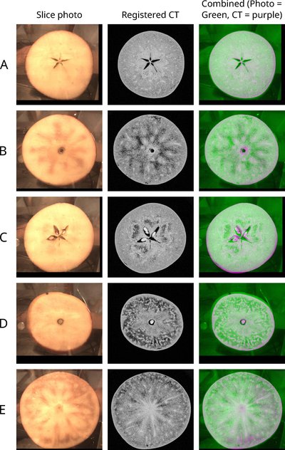
"Goudreinnetten, Kanzi's, Braeburns, all added up, I scanned five hundred apples one by one," says PhD candidate Dirk Schut as if it's the most ordinary thing in the world. The aim of his research: to determine whether you can see if an apple is good or already shows brown spots from the inside. Companies that inspect the quality of food before it goes to the supermarket are very interested in these types of techniques.

One such company is GREEFA, an industrial partner in Schut's research, which is connected to the Computational Imaging group of CWI. GREEFA makes sorting machines for fruits and vegetables. The sorting is done by using multiple parameters, such as the weight of the fruit and/or photographing it with different wavelengths, such as infrared light. By using image analysis, the bad specimens are filtered out. "The drawback is that you can only view the inside of an apple as a whole," says Schut. "On the CT scans that the FleX-ray creates, you can see every part of the apple in 3D. You can zoom in on any desired spot – for example, on an abnormal one – and see exactly where the discoloration stops."

Dirk Schut
Dirk Schut

1400 X-ray images

During the CT scan, the FleX-ray takes 1400 X-ray images of one apple from different angles. These images are then combined into a 3D model of the interior by using a mathematical algorithm. But that's not all, Schut explains. "We don't know what we're looking at: how do the different dents and spots look in an X-ray or CT scan? How do you distinguish a rotten spot from a slight discoloration?"

To answer that question, the PhD candidate sliced more than a hundred apples after scanning them and photographed those slices. Using a method he developed, he can find the correct region on the 3D CT scan for each photo of a piece of apple, so that you can compare both images side by side (see photo). If the slice has a rotten or brown spot, it immediately becomes clear how it looks on the scan.

Too slow

Schut also wanted to know how rotten or brown spots develop in apples in a storage. For this research he is working together with the university of Wageningen. To see how the CT images change over time, he scanned the same specimens at intervals several times. "Before they go to the supermarket, apples are stored in a low-oxygen environment," Schut explains. "As a result they remain fresh for a longer period. Most specimens look fine then. But in the first week after they come out of storage, brown spots develop the fastest. With the data I collect, I hope that a neural network can predict which apples will turn brown, so you can remove them in advance."

Although you can see much more on a CT scan, the scanning process still needs to be faster for the technique to be suitable for the food industry, where about eight apples per second pass by on a conveyor belt. "Each apple spends five minutes in the scanner here, which is way too slow. That's why we're investigating whether you can see the same brown spots in the individual X-ray images. While they may be less distinct due to lower contrast in X-ray images, they are captured in just one-tenth of a second."

Contact

Are you interested to learn more about our research, the FleX-ray Lab, or want to collaborate with us? CWI's Computational Imaging group can tell you all about this lab, contact them at seewhatsinside@cwi.nl

Timelapse of the scanning of an apple