Secrets of the skull

Our FleX-ray Lab houses a unique X-ray machine that creates 3D scans of the most diverse objects. This allows us to reveal details that remain hidden in regular scans. In this series, we showcase examples of what happens in the lab. Part 3: traces of malaria.

Publication date
17 Aug 2023

Did malaria, like the plague, have a significant impact on life in the Netherlands during the Middle Ages? There are strong indicators that it did. Osteoarchaeologist Rachel Schats from Leiden University is researching thousand-year-old skeletons for traces of this severe disease, which nowadays affects large portions of the population in tropical regions.

The skeletons with signs of this disease mostly come from the water-rich western Netherlands, where there were many marshlands at the time. These were ideal conditions for mosquitoes, the carriers of the malaria parasite. These parasites enter the body through mosquito bites, where they nest in red blood cells. This causes the cells to rupture, leading to anemia.
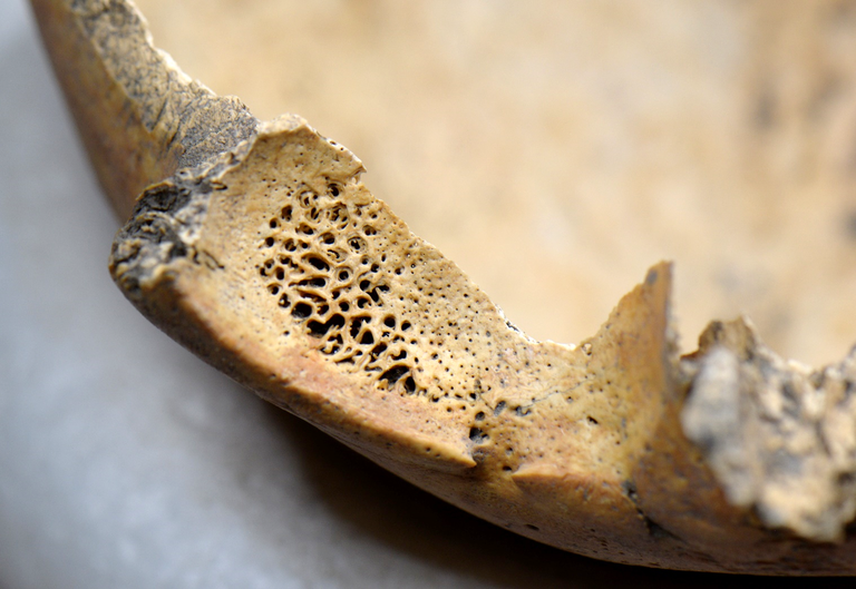
Holes

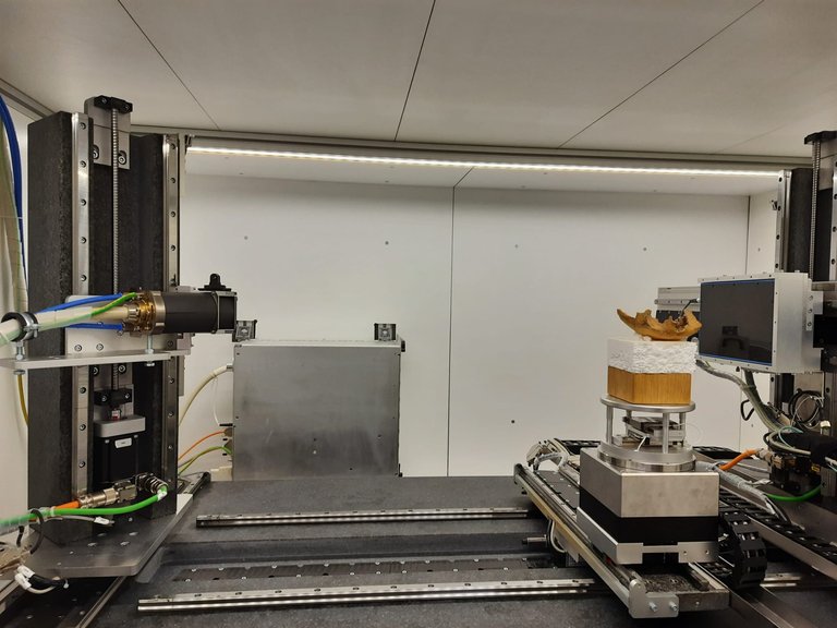
Anemia leaves traces on the bones, particularly in the eye sockets. Holes are formed there (see photo), known as cribra orbitalia. Schats is searching for these holes to gain an understanding of the extent and distribution of malaria in the Netherlands. However, cribra orbitalia can have other causes as well. How do you determine if a skeleton actually had anemia? "With the naked eye, I can't assess that with a hundred percent certainty," says Schats. This is how the researcher ended up at CWI, where nine eye sockets were scanned in the FleX-ray Lab.

Close up of a skull fragment with cribra orbitalia (tiny holes)
Cribra orbitalia (tiny holes) in part of an eye socket.

Schats wanted to examine the internal bone structure, the inside of the bone, so to speak. This structure looks different in cases of anemia compared to other conditions. During the scanning process, it was possible to zoom in on the inner part of the bone that forms the eye sockets. This allowed the Leiden researcher to have a 3D view of the degree of porosity (the number of small holes in the bone).

Many scans in a short time

One of the challenges in scanning the eye sockets was configuring the CT scanner. The settings had to be exactly the same for each specimen. CWI researcher Francien Bossema explains: "That was necessary to be able to compare the results properly. Due to the high resolution we aimed for in order to make the small holes clearly visible, we had to zoom in significantly on the eye sockets. This required two scans for a portion of the skull fragments, which were later digitally stitched together. So we had to make a lot of scans. And it all had to be done in one day." "From an ethical standpoint, we thought it best not to leave the skull fragments at CWI," Schats adds.

skull fragment inside the scanner
Skull fragment inside the FleX-ray scanner

Publication

"The scanning has proven to be very valuable," says Schats. "In most cases, there are indeed indications of anemia, even if the holes look different on the outside. In 2022, I presented the results at a conference, and I am currently writing a paper."

What does Schats ultimately hope to achieve? "That we get a better picture of health in this region during the Middle Ages. When people think of the Middle Ages, they immediately think of the plague. However, we don't have a clear picture of other diseases because there are too few written sources from that time. It's possible that malaria was truly a chronic disease with significant societal and economic impact."

Contact

Are you interested to learn more about our research, the FleX-ray Lab, or want to collaborate with us? CWI's Computational Imaging group can tell you all about this lab, contact them at seewhatsinside@cwi.nl

Inside the bone structure of an eye socket