Using Diffusion Models for Tomographic Imaging

We are looking for students who have a strong background in computational imaging or a related field, while a basic understanding of probability, inference, and generative AI is important. Familiarity in Python and deep learning frameworks such as PyTorch is also required.

Project description

Diffusion models have recently revolutionized image generation and are increasingly used as learned priors to recover missing information in inverse imaging problems. Computed Tomography (CT) reconstruction is one such problem, where information loss can occur due to limited measurements, motion, or dose constraints. Applying diffusion models to CT is a promising but non-trivial task, as these models usually require large training datasets, the pretrained models have mostly been trained on natural images or videos, and combining image generation with the physics of CT is not always straightforward. These open several interesting research directions.
In this project, you will choose from a few possible research directions: leveraging off-the-shelf video diffusion models for dynamic CT, noise schedule design for balancing learned prior and physics, or developing diffusion bridges that mimic the progressive CT acquisition process. Other related ideas on these topics are also welcome to discuss. Depending on the chosen direction, the work will either show how existing diffusion models can be leveraged for tomography without large CT datasets, or propose alternative strategies to achieve high-quality reconstructions under limited data conditions.

Supervision & focus areas

Supervisor : Tristan van Leeuwen (CWI),

Keywords : tomographic imaging, inverse problems, diffusion models
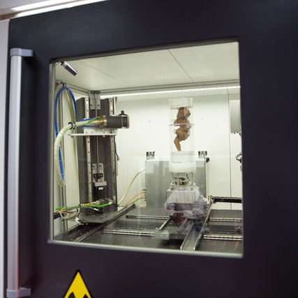
Secrets of the skull

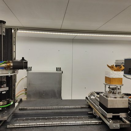
Our FleX-ray Lab houses a unique X-ray machine that creates 3D scans of the most diverse objects. This allows us to reveal details that remain hidden in regular scans. In this series, …

image

A Greek Mystery

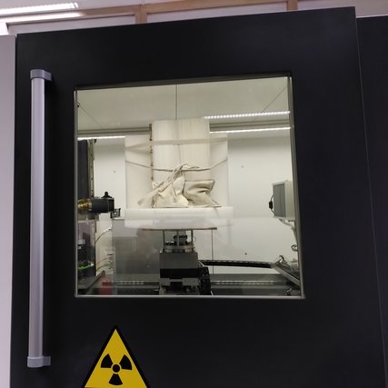
Our FleX-ray Lab houses a unique X-ray machine that creates 3D scans of a wide variety of objects. This reveals details that remain hidden in normal scans. In this series, we showcase …

IMG_20220426_093912

Looking for bad apples

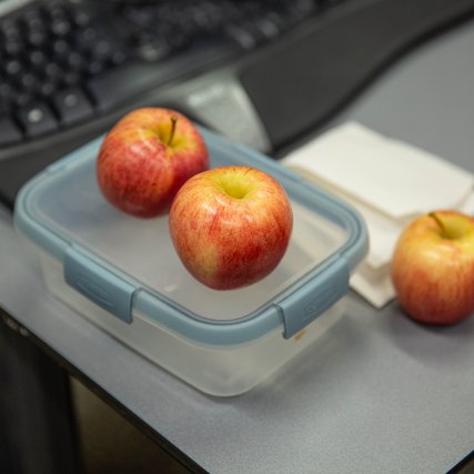
Our FleX-ray Lab houses a unique X-ray machine that creates 3D scans of the most diverse objects. This allows us to reveal details that remain hidden in regular scans. In this series, …

MK3_4751

NWO VIDI grant for Felix Lucka

X-ray Computed Tomography and AI team up to reveal fast dynamic processes.

CWI algemeen 2022 09 06-120

Towards no more glass in the jam with better X-ray scanners

X-ray and CT scanners are widely used devices in research, diagnostics and the industrial sector. And yet they are not nearly as fast and accurate as we would like. Mathé Zeegers is …

CI Flex lab

CONcISE Project aims for a revolution in biomedical optical imaging

The Computational Imaging (CI) group at CWI takes part in a new project funded by the European Union's Marie Skłodowska-Curie Actions (MSCA) of the Horizon Europe programme: CONcISE.

concise

IntACT: visualising the interior of art objects

The IntACT research team (CWI/Rijksmuseum) developed a tool to investigate art objects on the inside. In a new video the team explains the scope and outcome of the project.

Computational Imaging group and Rijksmuseum project awarded with NICAS funding. Image: Shutterstock.

Uncovering hidden features inside art objects in an interactive environment

In the past years, X-ray Computed Tomography (CT) has been used to visualise the interior of objects in 3D. For example within the NICAS and NWO funded Impact4Art project by CWI and …

CWI algemeen 2022 09 06-139