The FleX-ray Lab
The FleX-ray Lab embodies our vision on real-time, adaptive imaging. By transforming the linear imaging pipeline to a circular one, feedback from the expert can be incorporated directly in the scanning process. By coupling a versatile micro-CT scanner with high-end computational facilities, we can bridge the gap between theory and practice. Our scanner allows us to emulate various scanning geometries, including conveyor belt setups, and modern medical (helical) scans.
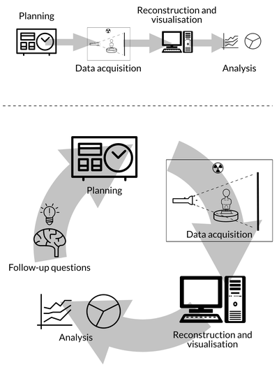
Figure 1: Transforming the linear imaging pipeline to a circular one, where the expert can be involved in the imaging in real time.
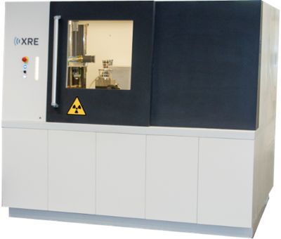
Figure 2: Our in-house microCT scanner at CWI.
Software
The ASTRA Toolbox is a MATLAB and Python platform providing scalable, high-performance GPU primitives for 2D and 3D tomography, including building blocks for advanced reconstruction algorithms. One of its main design goals is geometric flexibility, allowing the toolbox to be used with many types of experimental setups. The ASTRA Toolbox has been developed jointly by the Computational Imaging group of CWI, and the University of Antwerp. It is used in tomographic imaging research by academics working on novel algorithms and in experimental imaging labs. It is also deployed by several industrial partners as the core of the reconstruction software in their imaging equipment and applications. Furthermore, the toolbox is a main component in a number of other applications, including software used at two of the largest synchrotrons in the world: the European Synchrotron Radiation Facility (ESRF) in Grenoble, and the Advanced Photon Source (APS) in Chicago.
Tomosipo is a pythonic wrapper for the ASTRA-toolbox of high-performance GPU primitives for 3D tomography. The aim of tomosipo is to: expose a user-friendly API for 3D tomography, enable easy manipulation and visualization of 3D geometries, and express high-performance GPU-accelerated algorithms succinctly. Tomosipo substantially simplifies and accelerates the exploration of potential tomography setups, as well as the implementation of novel reconstruction algorithms. Moreover, tomosipo is especially useful when paired with neural networks, where fast GPU compute and easy integration with state of the art deep learning tools are essential.
The RECAST3D framework provides a real-time tomographic reconstruction and visualization pipeline for arbitrarily oriented 2D slices in a 3D volume. By reconstructing only the parts of the 3D volume requested by the user on demand, RECAST3D avoids the high computational cost of full 3D tomographic reconstructions. This lets the user effectively explore the 3D volume in real-time. It has been developed as a distributed, modular framework. This allows it to be used on individual workstations in lab settings, but also to be integrated into processing pipelines at large-scale experimental facilities such as synchrotrons. For example, it has been integrated into the high-speed imaging GigaFRoST system at the Swiss Light Source synchrotron. RECAST3D is GPU-accelerated by employing the ASTRA Toolbox, also jointly developed at CWI.
Research Themes
- Quantitative imaging is needed to fully exploit the potential of 3D imaging in many applications. To obtain reliable estimates of physical parameters (e.g., density or attenuation), more detailed mathematical models of the imaging system and proper calibration thereof are needed. In some cases this may lead to a non-linear inverse problem. A challenge here is to include proper calibration of the model to the imaging system. Furthermore, an estimate of the physical quantity of interest is useless without having a proper quantification of the uncertainty. While such uncertainty estimates are theoretically well-understood, naive (sampling) methods are intractable for large-scale problems.
- Multi-dimensional imaging entails both dynamic (time-resolved), and multi-parameter imaging (e.g., spectral or tensor imaging). The overarching challenge is that the image typically contains more parameters than can be estimated from the measurements. Prior information comes in the form of dynamical systems or structural assumptions about the object. Taking these in to account in the image reconstruction is computationally challenging, while identifying appropriate prior assumptions requires a data-driven approach.
- Non-linear models play an increasing role in many advanced imaging modalities. Moving away from linear models allows us to extract more information from the data but makes the image reconstruction a lot harder. Theoretical guarantees on recovery are much weaker and the algorithms are computationally much more expensive. To effectively deal with such non-linear imaging problems, novel methods are needed to mitigate the non-linearity and provide more robust image reconstruction methods. In particular, data-driven approaches are needed for proper calibration of non-linear models.
- High Performance Computing is a key ingredient when solving large-scale problems arising in practice. In particular, distributed GPU computing offers opportunities for many imaging and machine learning tasks. Development of practical tools for integrating various image reconstruction tasks with machine learning libraries are needed as well.
Applications
- Industrial quality control. Imaging plays an increasing role in Industry 4.0, where it aids in reducing waste and making production processes more sustainable. In the food processing and sorting industry, for example, X-ray imaging shows great potential for individualized quality control. To bring advanced CT techniques to the factory floor, several challenges need to be addressed; Fast 3D scanning and reconstruction is needed to meet throughput demand. Furthermore, fast and robust feature detection is needed to make decisions in real time. The Utopia project is a prime example of how interdisciplinary research can contribute to industrial and societal problems.
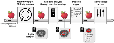
Figure 3: In the Utopia project, computational imaging is used for quality control of individual items in a sorting and processing line.
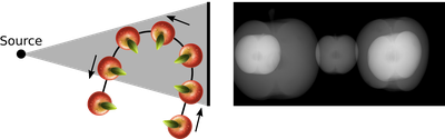
Figure 4: We develop novel imaging solutions for use in conveyor belt settings.
- Medical imaging. The demand for advanced diagnostic imaging is growing in order to maintain and improve health care worldwide. We develop novel image reconstruction algorithms for well established imaging techniques such as X-ray CT or MRI with the aim to overcome some of their disadvantages or limitations. For example, they can help to reduce the radiation dose in X-ray CT or lower the cost of an MRI examination, which will make it more accessible to patients worldwide. Robust data-driven methods based on machine learning are key components in these developments and topic of many of our new research lines. For certain diseases, novel imaging techniques that can provide more specificity and access to new diagnostic information are needed. For instance, we are developing Photoacoustic Tomography and Ultrasound Tomography for breast cancer screening and diagnosis.
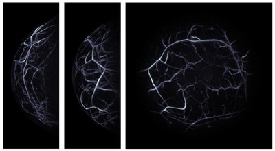
Figure 5: In Photoacoustic Tomography, ultrasound waves are induced by illuminating tissue with near-infrared light. From the resulting data, a three-dimensional reconstruction showing mainly the blood vessels inside the tissue can be made. In this example, this reconstruction is used for diagnoses and treatment planning of breast cancer.
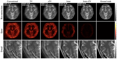
Figure 6: Magnetic Resonance Imaging is used for diagnosis and treatment planning for many diseases. If a patient moves while being scanned, this leads to blurring in the image. By incorporating a motion-estimation step in the image reconstruction, we get much sharper images.
- Cultural heritage. Most objects in the collections of cultural history museums can only be observed from the outside. However, the inside of the objects, which we cannot see, often contains a wealth of information about the making of the objects and their condition. Using 3D scanning techniques, such as CT scanning, these hidden traces can be visualized: finger prints of the maker that can be seen inside the clay, tool marks that tell us about the tools that have been used to craft the object, year rings inside wooden panels that can be used for dating, etc. The standard 3D scanning techniques have never been developed with these applications in mind, and often yield poor image quality when applied to cultural heritage objects. We develop novel algorithms and software that will enable us to create accurate 3D images of a broad range of museum objects, making scans within the walls of the museum and involving the cultural heritage experts. In the IMPACT4Art project, for example, we closely collaborate with experts from the Rijksmuseum.
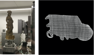
Figure 7: Dendrochronology can be used to determine the age and provenance of wooden art objects. We have developed CT techniques for non-destructive dendrochronology.
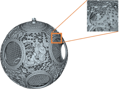
Figure 8: CT can give detailed insight in how objects were made. This 3D model, for example, revealed toolmarks that can be used to discover how these Chinese puzzle balls were made.
Contact
Are you interested to learn more about our research, the FleX-ray Lab, or want to collaborate with us? Feel free to contact our group leader, Tristan van Leeuwen at T.van.Leeuwen@cwi.nl.
More information
For recent news and, for instance, our group members, see our main page.